Gut microbiota and thyroid function

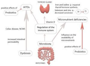
The gut microbiota plays a pivotal role in thyroid disorders, including Hashimoto thyroiditis, Graves’ disease and thyroid cancer. Recent data have suggested that microbes influence thyroid hormone levels through the regulation of iodine uptake, degradation, and enterohepatic cycling.

Studies have been shown that Lactobacillaceae and Bifidobacteriaceae are often reduced in hypothyroidism and hyperthyroidism. In Graves’ disease patients, the composition of the gut microbiota differs from that of healthy individuals, with a significant decrease in the relative abundance of Faecalibacterium prausnitziiButyricimonas faecalisBifidobacterium adolescentis and Akkermansia muciniphila compared to the control. Furthermore, a diagnostic model was developed using metagenome-assembled genomes of the gut microbiome, which may serve as a valuable predictor for Graves’ disease. The gut microbiota has the capacity to produce various neurotransmitters, such as dopamine, which can regulate the hypothalamus-pituitary axis and inhibit thyroid-stimulating hormone (TSH).

The Effects of Probiotics and prebiotics on thyroid function

 Numerous studies have been conducted to investigate the modulation of gut microbiota in order to restore dysbiosis in patients with thyroid disorders. Probiotics and prebiotics have demonstrated beneficial effects on thyroid diseases. Probiotics including bifidobacterium longum supplied with methimazole was implemented in nine patients with Graves’ disease for six months. The results showed a significant reduction in clinical thyroid indexes, including free T3, free T4, and thyrotropin receptor antibody (TRAb), while TSH levels increased compared to baseline. Another study explored the effects of a four-week treatment with a complex probiotics preparation consisting of Bifidobacterium infantisLactobacillus acidophilusEnterococcus faecalis and Bacillus cereus in patients post-thyroid hormone withdrawal (THW) following thyroid cancer surgery. The treatment led to a decreased occurrence of complications such as dyslipidemia and constipation. However, the serum levels of free T4, free T3 and TSH showed no change between groups. Synbiotics, which are a mixture of probiotics and prebiotics, have also been examined. They have shown potential in reducing TSH and increasing freeT3 in patients with hypothyroidism. A strategy based on microbiome-directed therapies, involving supplementation with probiotics, prebiotics and synbiotics holds promise as a therapeutic approach for thyroid disorders.

Conclusion

There is a growing interest in investigating the potential role of probiotics or prebiotics supplementation to improve thyroid function in humans. A recent meta-analysis indicates that supplementation with probiotics/prebiotics has no significant effect on thyroid hormone levels, while showing a modest decrease in TRAb levels. However, the results have been inconsistent, and the probiotics or prebiotics are various in each randomized clinical trial, leading a mixed effect.

References

Provided by: Dr. Nazila Kassaian

Techniques employed to explore and understand the Heart–Gut Axis

A New Approach to Gut–heart axis 

The human intestinal flora is closely related to human health. Gut microbiota dysbiosis is involved in the occurrence and development of various diseases, including coronary heart disease, hypertension, diabetes, inflammatory bowel diseases, and a wide range of inflammatory, metabolic, and neurological disorders. Studies in the last decade have proved that gut microbiota dysbiosis plays a crucial part in host inflammation and the formation of atherosclerosis and hypertension.

Challenges and future perspectives

It is difficult to apply what is known about microbiome composition and function in therapeutic settings. The dynamic nature of the microbiome, particularly it changes throughout illness development and in response to numerous variables such as nutrition, lifestyle, and pharmaceutical therapies, must be understood in order to address this, which calls for longitudinal investigations. Deeper research projects employing cutting-edge approaches like next-generation sequencing, metatranscriptomics, metaproteomics, and metabolomics, as well as computational methodologies, are currently of the utmost importance.۴ We can now investigate the complex interactions of the Heart–Gut axis thanks to these instruments. We will be able to identify the processes by which the gut microbiota affects medication metabolism and response by merging functional analysis, simultaneous profiling of the gut microbiome, metabolomics, and other omics data. These multifaceted techniques have a great deal of promise for unravelling the complex mechanisms by which the gut microbiota interacts with the host, ultimately revealing insightful information for enhancing medication, therapy and enhancing patient outcomes.

Targeting gut microbiota as a therapeutic option

  • There is interest in employing microbiome-based therapeutics as a possible therapeutic option for CVDs given the potential impact of the gut microbiota on cardiovascular health. Probiotics, which are living bacteria that provide health advantages when taken in sufficient quantities, are one strategy. Numerous beneficial bacterial strains like  bifidum, L. casei, L. Acidophilus, L. zisttakhmir, L. reuteri, L.fermentum, C. butyricum, B.breve,have been tested for the treatment of CVDs both in animal models and humans.
  • Prebiotics and CVDs study is still in its infancy, but there is mounting evidence that they may have beneficial effects on several risk factors for cardiovascular disease like-blood pressure, cholesterol management, chronic inflammation, oxidative stress, glucose metabolism, and weight management. However, further studies are required to evaluate the effectiveness of these probiotics and the best dosage.
  • Another method is fecal microbiota transplantation (FMT), which involves inserting healthy donor’s feces into the recipient’s gut. Although research is still being done to determine FMT’s potential therapeutic function in CVDs, it has historically been utilized largely to treat illnesses like Clostridium difficile Following FMT, preliminary research in animal models has demonstrated encouraging improvements in cardiovascular parameters and atherosclerotic reduction. The use of gut microbiota-based therapeutics for CVDs is still in its infancy, and more study is required to completely comprehend the intricate relationships between the gut and the heart and to assess the efficacy and safety of these strategies. Individual differences in gut microbial makeup and responsiveness to therapies may also provide challenges in developing personalized therapy modules and regimen.
  • Research has shown AST-120, an oral charcoal adsorbent to have beneficial effect in the treatment of CVDs. However, it is unclear how adsorbents may affect the gut microbiota’s composition and its associated metabolites in people with CVDs.

Reference

Majumder S, Kirtikumar RM, Shetty V, Mukherjee S, Narayan P. Cardiovascular diseases and the heart–gut cross talk. Indian Heart Journal. 2023 Dec 7.

Provided by: Dr. Nazila Kassaian

The contribution of the gut microbiome to bariatric surgery

The gut microbiome has been identified as a potential factor in weight regulation. Thirty-nine percent of the adults are overweight (25–۲۹.۹ kg/m2) and 13% are obese (BMI≥۳۰kg/m2) worldwide. Recently, it has been found that obesity may affect brain function and structure, as it is associated with impaired cognition and alterations in gray matter (GM) and white matter (WM). Moreover, a higher BMI and waist-to-hip ratio (WHR) have been associated with lower fractional anisotropy (FA) values. Moreover, it is proposed that obesity increases the risk of developing dementia later in life by 60–۹۰%, versus healthy weight individuals. A growing of evidence reveals that obesity is related with alterations in neuroendocrine production and secretion, including ghrelin, insulin, GLP-1 and PYY.

Bariatric surgery for obesity treatment

Bariatric surgery is an effective treatment for obesity leading to rapid and sustainable weight loss. Bariatric surgery decreases body weight not only due to physical effects such as reduced food intake and malabsorption but also due to the various neuroendocrine changes which affect energy homeostasis and hunger/satiety. Moreover, Bariatric surgery might improve the gut microbiota diversity and restore white adipose tissue function, which can improve obesity-related immunological and cognitive impairments. The gut-brain axis consists of a bidirectional communication system, connected through the vague nerve, spinal fibers and sympathetic and parasympathetic fibers which are directly innervating the gastrointestinal tract. These elements communicate through endocrine messengers, neuro-immune mediators and neuroactive metabolites.

Conclusion

To summarize, Bariatric surgery is a good procedure to treat obesity and its related pathologies, however long-term effects remain unsolved. Future research should focus on the long-term effects of Bariatric surgery, to be able to investigate the neuroendocrine, microbiota and white adipose tissue changes and to potentially determine the new “normal” after homeostatic adjustments. Various studies have focused on neuroendocrine alterations already after six months. Six months post-surgery patients lose weight rapidly and generally still follow their post-operative diet. Therefore, the observed effects 6 months post-surgery might differ at longer follow-ups, when patients achieve a stable weight, or regain weight.

References

Provided by: Dr. Nazila Kassaian

درمان سلیاک با اصلاح میکروبهای روده

سلیاک یک بیماری شدید خود ایمنی است. در این بیماری، سلولهای ایمنی، روده کوچک را مورد حمله قرار می دهد. سلیاک حساسیت شایعی است که در افرادی با برخی از ژن‌ها هنگامی که گلوتن را مصرف می‌کنند رخ می‌دهد. گلوتن در غلاتی مانند گندم، جو و چاودار وجود دارد. این افراد بدنبال مصرف این غذاها دچار سوء جذب و دل درد می شوند. اخیرا ارتباط سلیاک با میکروبیوتای روده کشف شده است. میکروبیوتا، گروهی از میکروارگانیسم‌ها هستند که در روده ما زندگی می‌کنند و تعادل و تنوع آنها ضامن سلامتی بدن است.

مقایسه میکروبیوتای افراد سالم با بیماران سلیاکی

تحقیقات نشان می‌دهد ترکیب میکروبیوتا در افراد مبتلا به بیماری سلیاک با افرادی که این بیماری را ندارند، متفاوت است. در واقع، بیماران مبتلا به بیماری سلیاک در مقایسه با افراد سالم دارای کاهش در گونه های مفید و افزایش در گونه های بالقوه بیماری زا هستند. این دیس بیوز ممکن است پس از یک رژیم غذایی بدون گلوتن همچنان باقی بماند. بنابراین، میکروبیوتای روده می تواند نقش مهمی در پاتوژنز بیماری سلیاک ایفا کند. برخی از مطالعات نشان می‌دهند که ترکیب میکروب‌ها در بدن افراد مبتلا به بیماری سلیاک، به شکلی است که با هضم گلوتن  باعث افزایش آسیب‌پذیری در برابر بیماری می شود.  از طرف دیگر، میکروب‌ها در هضم سایر مواد غذایی نیز دخیل هستند و تغییراتی در این فرآیند در افراد مبتلا به بیماری سلیاک مشاهده شده است.  به طور کلی، میکروبیوتا می‌تواند به عنوان یک عامل همراه، بسته به فعالیت و نوع میکروب‌های موجود در دستگاه گوارش، منجر به افزایش یا کاهش آسیب‌پذیری در برابر بیماری سلیاک شود. اما قطعا باید ژن‌های مربوطه و تماس با گلوتن نیز وجود داشته باشد.

Celiac

باید توجه نمود که تبعیت از رژیم غذایی بدون گلوتن در صورتی که بدون نظارت و تجویز متخصص انجام ‌شود، می‌تواند منجر به کمبود عناصر مغذی و مشکلات دیگر شود. لذا، تشخیص درست بیماری سلیاک و بدنبال آن رژیم غذایی بدون گلوتن از یک طرف و تلاش در جهت اصلاح فلور میکروبی روده از طریق مصرف فیبرها و پروبیوتیکها از طرف دیگر برای مدیریت و بهبود این بیماری بسیار مهم است. به نظر می رسد استفاده از پروبیوتیک ها پاسخ التهابی را کاهش داده و نسبت طبیعی باکتری های مفید را در دستگاه گوارش بازیابی می کند. برای درک بهتر نقش میکروبیوتای روده در پاتوژنز بیماری سلیاک و تأثیر بالینی و استفاده درمانی پروبیوتیک ها در این زمینه، به شواهد بیشتری نیاز است.

مترجم: راحم رحمتی (Rahem Rahmati)

ویراستار:نازیلا کسائیان (Nazila Kassaian)

References

Pecora F, Persico F, Gismondi P, Fornaroli F, Iuliano S, De’Angelis GL, Esposito S. Gut microbiota in celiac disease: is there any role for probiotics? Frontiers in Immunology. 2020 May 15; 11:957.

Chibbar R, Dieleman LA. The gut microbiota in celiac disease and probiotics. Nutrients. 2019 Oct 5; 11(10):2375.


بایدها و نبایدهای تغذیه ای در بیماران IBD

IBD

بایدها و نبایدهای غذایی در بیماریهای التهابی روده (IBD) یکی از سوالات اصلی بیماران پس از تشخیص است. اگرچه هیچ غذا یا رژیم غذایی خاصی وجود ندارد که کاملا بتواند از کولیت اولسراتیو و بیماری کرون پیشگیری و یا آنها را درمان کند، تغذیه و رژیم غذایی مناسب می تواند به بهبود علائم و کاهش التهاب روده کمک کند. نشان داده شده است که مصرف سبزیجات، میوه، آجیل، حبوبات، روغن زیتون و منابع پروتئینی بدون چربی مثل لوبیای سویا، گوشت مرغ و ماهی، اثر محافظتی در برابر ابتلا به IBD دارد و به بهبود جمعیت میکروبی روده کمک می کند. در مقابل، الگوهای غذایی غربی، سرشار از اسیدهای چرب غیراشباع امگا ۶، الکل، گوشت قرمز و افزودنی‌های غذایی (نمک بیش از حد و شیرین‌کننده‌های مصنوعی) التهاب روده را شدت بخشیده و می‌توانند علائم این بیماری را بدتر کنند. به گفته محققین، رژیم غذایی غنی از اسیدهای چرب غیراشباع امگا ۶ (که معمولاً در روغن‌های ذرت، سویا، گلرنگ و آفتابگردان یافت می‌شود) باعث التهاب روده می‌شود. در مقابل، رژیم غذایی غنی از روغن زیتون و حاوی اسیدهای چرب غیراشباع امگا ۳ (روغن ماهی) و چربی لبنیات باعث تقویت ایمنی در کولیت اولسراتیو می شود.

تغذیه درمانی در شرایط حاد بیماری

در شرایط حاد، بهترین رژیم درمانی ، تغذیه انترال (EEN) است که یک فرمولای مایع است. در این رژیم تمام مواد مغذی ضروری تامین می شود و هیچ غذای دیگری داده نمی شود. این رژیم بخصوص برای کاهش التهاب کودکان موثر است.. در نهایت، EEN باید برای مدت زمان محدودی استفاده شده و سپس غذا به تدریج اضافه می شود.

نتیجه گیری

امروزه رژیم غذایی مدیترانه ای برای افراد مبتلا به IBD که به دنبال یک رژیم غذایی متعادل و سالم هستند توصیه می شود.دانشمندان معتقدند تغذیه و نوع رژیم غذایی افراد IBD باید با توجه به شدت بیماری و آنچه در سبک زندگی آنها اتفاق می افتد، شخصی سازی شود.  بنابراین مشاوره با یک متخصص تغذیه با تخصص در مدیریت IBD برای ایجاد یک برنامه فردی ضروری است. توصیه می کنیم قبل از انتخاب یکی از رژیم‌های غذایی مد روز که بعضا بصورت آنلاین تبلیغ می‌شوند، با پزشک خود صحبت کنید تا شما را به یک متخصص تغذیه وارد در این بیماری معرفی کند و یک برنامه غذایی شخصی‌سازی شده برای شما تجویز شود.

مترجم: راحم رحمتی(Rahem Rahmati)- نازیلا کسائیان(Nazila Kassaian)

ویراستار: مرضیه رحیم خراسانی (Marzieh Rahim Khorasan) – پریسا هاشمی ( Parisa Hashemi)


References

ارتباط ژن های مقاومت آنتی بیوتیکی نوزاد با میکروبیوتای روده و شیر مادر

اثرات آنتی بیوتیک ها بر تنوع باکتریایی روده انسان در اوایل زندگی از اهمیت ویژه ای برخوردار است. مشخص شده است که روده نوزادان بدون توجه به قرار گرفتن در معرض آنتی بیوتیک، مقادیر بالایی از ژن های مقاوم به آنتی بیوتیک را در مقایسه با بزرگسالان حمل می کند.

مطالعه جدید نشان می دهد، میکروبیوم‌های روده و شیر مادر ممکن است در دستیابی نوزاد به ژن‌های مقاومت آنتی‌بیوتیکی و عناصر ژنتیکی متحرک نقش داشته باشند. محققان شیر مادر و میکروبیوم های مدفوع نوزاد و مادر را طی ۸ ماه آنالیز کردند. میکروبیوتای مدفوع نوزادان، ژن‌های مقاومت آنتی‌بیوتیکی و ترکیبات ژنتیکی متحرک نوزادان بسیار شبیه ترکیبات مادرانشان بود. بالاترین سطوح شباهت مشترک در شیر مادر مشاهده شد، به طوری که ۷۰ درصد از ژن های مقاومت آنتی بیوتیکی در شیر مادر با ژن های موجود در روده نوزاد همپوشانی داشتند.

همچنین نوزادان مادرانی که در حین زایمان آنتی‌بیوتیک دریافت کرده‌اند، دارای تعداد بیشتری از ژن‌های مقاومت آنتی‌بیوتیکی خاص و همچنین عناصر ژنتیکی متحرک بودند. علاوه بر این، تغذیه با شیر مادر برای کمتر از ۶ ماه منجر به افزایش برخی از ژن های مقاومت آنتی بیوتیکی مانندتتراسایکلین ها بود.

فراوانی ژن مقاومت آنتی بیوتیکی نیز با ترکیب میکروبیوم نوزادان و مادران مرتبط بود. این نتایج نشان می دهد که برهمکنش بین گروه های مختلف طبقه بندی در میکروبیوم روده نوزادان و مادران و در شیر مادر ممکن است مقاومت آنتی بیوتیکی را شکل دهد. اشرشیا کلی باکتری مشترکی بود که به شدت با فراوانی ژن های مقاومت آنتی بیوتیکی همبستگی داشت.

نتیجه گیری

یافته‌ها نشان می‌دهند، ژن مقاومت آنتی بیوتیکی هم از طریق شیر مادر و هم از طریق میکروبیوتای روده مادر منتقل می شود. با این وجود، ترکیب میکروبیوم روده تعیین کننده اصلی در بار مقاومت کلی آنتی بیوتیک است.

مترجم: پریسا هاشمی (Parisa Hashemi)

ویراستار: مرضیه رحیم خراسانی (Marzieh Rahim khorasani)

Reference

https://www.gutmicrobiotaforhealth.com/the-source-of-antibiotic-resistance-genes-in-the-infant-gut-may-rely-on-the-mothers-gut-and-breast-milk-microbiota/?search=antibiotic


Some Vitamins Categorized as a Prebiotic

The International Scientific Association for Probiotics and Prebiotics (ISAPP) has been redefined prebiotics in 2017. They suggested that some vitamins were included in the category of prebiotics. Vitamins, as organic components, are usually present in foods instead of synthesized in sufficient quantities by the host and can play fundamental role in mediating biological processes in microbes, maintaining microbial homeostasis and intestinal barrier integrity and so, may have a potential use as prebiotics. Vitamins can be divided into two categories: lipophilic vitamins; consisting of vitamin A, D, E and K, and hydrophilic vitamins, which include vitamin B and C.

The effects of fat soluble vitamins on microbiota

Vitamin D is one of the most studied vitamins in the context of gut microecology. Some studies have demonstrated that the composition and diversity of gut microbes are strongly affected by vitamin D deficiency. For example, in an interventional, open-label pilot study, vitamin D3 supplementation was found to decrease the relative abundance of Proteobacteria while increasing that of Bacteroidetes (1). Furthermore, Guida et al. (2) evaluated a diet deficient in vitamin D on a mice model and found thatthe abundance of Firmicutes, Verrucomicrobia and Bacteroidetes decreased. In addition to vitamin D, oral administration of some other vitamins has also been shown to affect the composition of gut microbes.
Tian et al. analyzed fecal samples from vitamin A sufficient and deficient mice and observed a higher
Firmicutes/Bacteroidetes (F/B) ratio in these mice, which is associated with various metabolic diseases (3).
On the contrary, another study found that feeding mice with low levels of vitamin E increased the
abundance of Firmicutes, resulting in a higher F/B ratio in the gut, whereas no significant differences were observed in mouse fed with high doses of vitamin E as compared to the control group (4). Vitamin K in human body usually taken from dietary supplements. Gut microbiota also possess the gene to synthesize vitamin K (5). Dietary vitamin K can be remolded by bacteria such as Eubacterium rectale, Bacillus subtilis and several Bacteroides species in the form of menaquinones, which are able to regulate gut microbiota. Since some genera of Bacteroides and Faecalibacterium lost the ability to synthesize menaquinones, they have to utilize menaquinones made by nearby bacteria as growth factors for themselves (6).

The effects of water soluble vitamins on microbiota

Vitamin B12 may make a contribution in shaping the structure and function of human gut microbial communities through altering the corrinoid profile (7). Oral vitamin B12 supplement may also selectively deplete Bacteroides in C57BL/6 mice (8). This difference suggests that different types of vitamins and different doses of the same vitamin may have different regulatory effects on gut microbes. Specific screening of vitamins and next generation of probiotics (NGPs) is necessary to establish the mechanism by which vitamins affect the gut microbiota in order to determine which vitamins can be used as prebiotics for which NGPs.
Vitamin B2 (riboflavin) supplementation is critical for maintaining the abundance of Faecalibacteriome. Prausnitzii (F. P) in the gut microbiota. Although F. P adheres to the gut mucosa where oxygen diffuses from epithelial cells, it can employ an extracellular electron shuttle of riboflavin and thiols to transfer electrons to oxygen (9).

Provided by: Dr. Nazila Kassaian

Edited by: Marzieh Rahim khorasani

References:


Quantitative PCR provides a simple and accessible method for quantitative microbiota profiling

Introduction

The use of relative abundance data from next generation sequencing (NGS) can lead to misinterpretations of microbial community structures, as the increase of one taxon leads to the concurrent decrease of the other(s) in compositional data. . Since the changes of components are mutually dependent, high false discovery rates occur when compositional data are analyzed using traditional statistical methods.  Although different DNA- and cell-based methods as well as statistical approaches have been developed to overcome the compositionality problem, and the biological relevance of absolute bacterial abundances has been demonstrated, the human microbiome research has not yet adopted these methods, likely due to feasibility issues. Here, we describe how quantitative PCR (qPCR) done in parallel to NGS library preparation provides an accurate estimation of absolute taxon abundances from NGS data and hence provides an attainable solution to compositionality in high-throughput microbiome analyses. The advantages and potential challenges of the method are also discussed.

Method

۱-Bacterial DNA extraction

Bacterial DNA will be extracted from fecal samples using a modified version of repeated bead beating that efficiently extracts bacterial DNA from both Gram-positive and -negative bacteria.

۲-۱۶S rRNA gene sequencing

۳-Sequencing data processing and analysis

The preprocessing will be done in the R package mare, utilizing USERACH for quality filtering, chimera removal, and taxonomic annotation. Only the high-quality forward reads should be used.

۴-Quantitative PCR

Quantification of total bacteria, specific taxa and butyrate production capacity should be carried out by qPCR.

۵-Calculation of absolute abundance and copy-number correction

The sequencing reads assigned to different taxa in each sample will be divided by the total number of reads for the sample to obtain relative abundances of the taxa in each sample. The relative abundances obtained based on the sequencing reads will be translated into total abundances by multiplying the relative abundance of each taxon by the total bacterial abundance in the sample. These figures will be further corrected for 16S rRNA gene copy-number variation by dividing the abundance of a taxon by the number of 16S copies in its genome. For the copy-number correction, the 16S copy number database rrnDB can be used.

PCR provides

Conclusion

Importantly, qPCR-based quantitative microbiome profiling enjoys the following conceptual and practical benefits over other approaches:

۱-Cost-effectiveness and feasibility: qPCR is cost-effective and accessible as the laboratory settings, machinery and reagents are similar to those needed for preparing the NGS libraries. The same DNA extract serves as the starting material both for qPCR and NGS, making qPCR done in 96- or 384-format easy to implement in the workflow for high-throughput analysis of up to thousands of microbiome samples.

۲-Simplicity: qPCR is relatively simple to perform compared to flow cytometry that requires considerable expertise for reproducible results. In fact, flow cytometric enumeration of microbial cells was initially restricted to pure cultures and still remains challenging when performed in complex matrices [32]. Also, no spikes, other exogenous controls, or complicated transformation/computation are needed in qPCR-based quantitative microbiome profiling.

۳-Comparability to NGS: Unlike flow cytometry that counts cells, qPCR and NGS both target bacterial DNA, including extracellular DNA derived from lysed bacteria. Extracellular DNA can be intrinsic or result from the differential lysis of Gram-positive and negative bacteria during the common freeze-thawing prior to fecal DNA extraction. As the 16S profiles from the gut appear very different for intracellular and extracellular DNA [33], qPCR is expected to reflect the NGS targeted community structure both quantitatively and qualitatively more closely than flow cytometry

۴-Applicability: qPCR-based quantitative microbiome profiling is applicable also for samples containing a substantial amount of host or non-bacterial DNA, in which bacterial density cannot be reliably estimated by total DNA yield [5]. Moreover, the qPCR-based method can be employed to study also non-bacterial communities where a universal marker gene is available, such as in fungi

References

Knight R, Vrbanac A, Taylor BC, Aksenov A, Callewaert C, Debelius J, et al. Best practices for analysing microbiomes. Nature reviews Microbiology. 2018; 16(7):410–۲۲. Epub 2018/05/26. https://doi.org/10. 1038/s41579-018-0029-9 PMID: 29795328.

Morton JT, Marotz C, Washburne A, Silverman J, Zaramela LS, Edlund A, et al. Establishing microbial composition measurement standards with reference frames. Nat Commun. 2019; 10(1):2719. Epub 2019/06/22. https://doi.org/10.1038/s41467-019-10656-5 PMID: 31222023; PubMed Central PMCID: PMC6586903.

Props R, Kerckhof FM, Rubbens P, De Vrieze J, Hernandez Sanabria E, Waegeman W, et al. Absolute quantification of microbial taxon abundances. The ISME journal. 2017; 11(2):584–۷. Epub 2016/09/10. https://doi.org/10.1038/ismej.2016.117 PMID: 27612291; PubMed Central PMCID: PMC5270559.

Jian C, Luukkonen P, Yki-Järvinen H, Salonen A, Korpela K. Quantitative PCR provides a simple and accessible method for quantitative microbiota profiling. PLoS One. 2020 Jan 15; 15(1):e0227285.

The role of the microbiota in the management of intensive care patients

The role of the microbiota in the management of intensive care patients

In the gut, the microbiota mostly comprised bacteria, but it also harbors archaea, viruses, protozoans, and fungi. The composition of the gut microbiota is unique to each individual in that the gut microbiota of two given individuals consistently show differences in their composition. Nonetheless, it is also highly dynamic and evolves throughout life under the influence of a wide diversity of genetic, environmental, medical, and dietary determinants.

The microbiota and intensive care patients

In the intensive care setting, the gut microbiota of patients is submitted to various stresses including antibiotic exposure, modification of gastrointestinal transit, artificial nutrition or sepsis which may lead to a dysbiosis during hospitalization. Indeed, the gut microbiota in critically ill patients appears to be different from that of healthy subjects, demonstrating markedly lower richness and diversity, and the near replacement of commensal genera by opportunistic pathogens. Recent evidence has shown that dysbiosis in ICU patients might have consequences on survival, stressing that dysbiosis could be considered as an authentic, organ-failure-affecting prognosis along with renal, cardiac, or respiratory failures.

Dysbiosis alteration and patients’ management

The gut microbiota mainly includes difficult-to-cultivate anaerobic bacteria, hence knowledge about its composition has significantly arisen from culture-independent methods based on next-generation sequencing (NGS) such as 16S profiling and shotgun metagenomics.

Correcting the microbiota disturbances to avoid their consequences is now possible. Fecal microbiota transplantation is recommended in recurrent C. difficile infections and microbiota-protecting treatments such as antibiotic inactivators are currently being developed. The growing interest in the microbiota and microbiota-associated therapies suggests that the control of the dysbiosis could be a key factor in the management of critically ill patients.

The gut microbiota is also the main reservoir for multidrug-resistant bacteria organisms (MDRO). Initially kept at low intestinal concentrations as a consequence of the barrier effect exerted by commensal anaerobic bacteria, they may bloom after antibiotic exposure and increase the risk their involvement in further infections.

prepared by: Nazila Kassaian

Reference

Szychowiak P, Villageois-Tran K, Patrier J, Timsit JF, Ruppé É. The role of the microbiota in the management of intensive care patients. Ann Intensive Care. 2022 Jan 5;12(1):3. doi: 10.1186/s13613-021-00976-5. PMID: 34985651; PMCID: PMC8728486.

?What is the microbiome

microbiome

microbiome is estimated that at least 100 trillion (10۱۴) microbial cells and millions of viruses reside within the human body, all of which produce various enzymes, chemicals, hormones, and vitamins that can interact with other human cells.

human microbiome

This complex community of microorganisms, which is more commonly referred to as the microbiota, can be of viral, bacteria, archaea, and/or eukaryotic etiologies. Comparatively, the genes that these organisms encode for is known as the microbiome. Gut microbiota (formerly called gut flora) is the name given today to the microbe population living in our intestines. It contains hundreds of trillions of microorganisms. One third of our gut microbiota is common to most of the people, while two thirds are specific to each one of us. In other words, the intestinal microbiota is a sort of individual identity card.

As a result of extensive research that has been conducted over the past few decades, microbial organisms can have significant impacts on the health of humans.

Moreover, the microbiome can determine how the immune system responds to potential pathogens, the rate at which nutrients and energy are absorbed from the diet as well as certain psychological and behavioral states.

When the balance between the gut microbiota and the host is disrupted, which is a condition otherwise referred to as dysbiosis, various health conditions can occur, some of which include malnutrition, inflammatory bowel diseases (IBD), obesity, neurological disorders, and cancer.

gut microbiome

Early diversification of the microbiome

During fetal development and infancy, the diversity of both the bacteria and viruses that comprise the microbiome is generally low. Since the gut, during these early stages of life, contains oxygen, the microorganisms that are present within the gut at this time are mostly aerotolerant.

As the child develops, these organisms are replaced by anaerobes. This evolution of microbial organisms within the gut is rapid, as studies have even shown that 56% of the virome sequences found within the gut of infants during their first week of life are not present after the second week.

This diversity continues to expand rapidly throughout the first 3 months of life, which is comparable to the virome of the adult microbiome, in which approximately 95% of these microorganisms are conserved over time.

Since virus particles are not present in either formula or breast milk, researchers believe that this early diversification of the microbiome is the result of environmental exposures and maternal contact.

 The steps that could preserve the current state of the gut microbiota

  • Using antibiotics wisely
  • Reducing the number of the unnecessary Cesarean sections
  • Promoting breastfeeding
  • Reducing antimicrobial products in our environment
  • Improving the diet by increasing the amount of fiber and diversity of foods to promote microbial diversity and benefit health
  • Adding functional foods containing pro-, pre- and postbiotics to diets

reference

Grieneisen L, Dasari M, Gould TJ, Björk JR, Grenier JC, Yotova V, Jansen D, Gottel N, Gordon JB, Learn NH, Gesquiere LR. Gut microbiome heritability is nearly universal but environmentally contingent. Science. 2021 Jul 9;373(6551):181-6.