میکروبیوتای واژن و ارتباط آن با میکروبیوم روده

در حالی که محبوب ترین میکروبیوتایی که ممکن است در مورد آن شنیده باشید میکروبیوتای روده است، واژن نیز میکروبیوتا دارد. در مورد رابطه بین این دو و اینکه چرا مراقبت از میکروبیوتای روده برای میکروبیوتای واژن اهمیت دارد بیشتر بدانید.

چرا میکروبیوتای واژن برای سلامتی مهم است؟

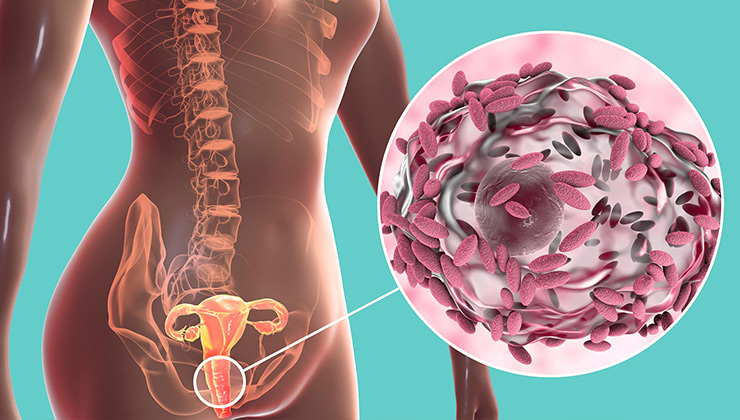
میکروارگانیسم ها در هر ناحیه ای از بدن که در معرض خارج قرار دارد وجود دارند: پوست، گوش، بینی، مجاری تنفسی و واژن. میکروبیوتای واژن از تعداد زیادی باکتری (عمدتاً گونه های لاکتوباسیلوس) و تعداد کمی قارچ (بیشتر کاندیدا آلبیکنس) تشکیل شده است. نکته مهم این است که برخلاف میکروبیوتای روده، میکروبیوتای واژن در شرایطی که تنوع آن کم باشد، وضعیت خوبی دارد. هنگامی که سایر باکتری ها (گاردنرلا واژینالیس، آتوپوبیوم واژن و دیگران) یا قارچ ها (گونه های کاندیدا) بیش از حد رشد می کنند و لاکتوباسیل ها کاهش می یابند، عفونت واژن می تواند ظاهر شود.

نقش میکروبیوتای واژن

  • تولید ترکیبات دفاعی، مانند اسید لاکتیک و مواد ضد میکروبی
  • عمل به عنوان یک مانع و تحریک تولید موکوس توسط اپیتلیوم واژن
  • حمایت از سیستم ایمنی بدن زنان

راهیابی میکروب های مضر از روده به واژن

انتقال باکتری از رکتوم به واژن یکی از چندین علت شناخته شده عفونت های واژن است. اگرچه روده انسان محلی برای میکروارگانیسم هایی است که به عنوان میکروبیوتای سالم روده شناخته می شوند، اما زمانی که به واژن سفر می کنند ممکن است مشکلاتی ایجاد کند. این موضوع در مورد برخی از باکتری ها مانند اشریشیا کلی و برخی قارچ ها مانند گونه های کاندیدا صدق می کند. هنگامی که این میکروارگانیسم‌ها از رکتوم به واژن حرکت کرده و در آن رشد می‌کنند سبب عدم تعادل میکروبیوتای واژن شده و منجر به عفونت آن می شود.

تجویز و استفاده از پروبیوتیک ها منجر به مهاجرت میکروب های مفید از روده به واژن می‌ شود و این امر بر سلامت واژن اثرگذار است. بنابراین اگر میکروبیوتای روده شما در وضعیت خوبی قرار داشته باشند، میکروبیوتای واژن شما نیز در حالت محافظتی قرار خواهند گرفت. میکروبیوتای روده نیز می توانند هورمون های استروژن را متابولیزه و به جای دفع از طریق مدفوع، منجر به تجمع آنها در بدن شود بنابراین افزایش استروژن در واژن باعث افزایش تولید گلیکوژن به عنوان منبع غذایی برای لاکتوباسیل های مفید می شود.

عدم تعادل میکروبیوم واژن منجر به واژینوز باکتریایی می شود

بسته به چرخه قاعدگی، رژیم غذایی، اقدامات بهداشتی، داروها، استرس و بیماری میکروبیوتای واژن هر زن متفاوت است. رشد بیش از حد میکروارگانیسم‌های بیماری‌زا در واژن می‌تواند در نیمی از زنان بدون علامت و یا همراه با ترشحات غلیظ و سفید با بوی ماهی گندیده، درد یا خارش همراه باشد.

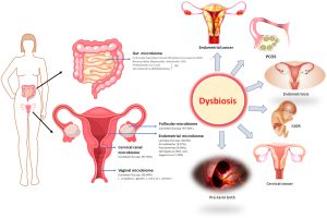
تغییر میکروبیوتای واژن یا دیس بیوز واژن با واژینوز باکتریایی، برفک، عفونت های مکرر دستگاه ادراری و حتی کاهش باروری همراه است. این یافته‌ها امکان اصلاح میکروبیوتای واژن را به‌طور مستقیم یا غیرمستقیم با تأثیر بر میکروبیوتای روده برای بهبود سلامت زنان باز می‌کند.

نتیجه گیری

پاتوژن های روده می توانند به داخل واژن رفته و به بر هم خوردن تعادل میکروبی واژن کمک کنند؛ بنابراین مراقبت از روده قطعا سلامت واژن را بهبود می بخشد.

مترجم: راحم رحمتی (Rahem Rahmati)، پریسا هاشمی ( Parisa Hashemi)

ویراستار: مرضیه رحیم خراسانی (Marzieh Rahim khorasani)

References

Bradford LL, Ravel J. The vaginal mycobiome: A contemporary perspective on fungi in women’s health and diseases. Virulence. 2017; 8(3):342-351. doi: 10.1080/21505594.2016.1237332.

Nair Balakrishnan S, Yamang H, Lorenz MC, et al. Role of vaginal mucosa, host immunity and microbiota in vulvovaginal candidiasis. Pathogens. 2022; 11(6):618. doi: 10.3390/pathogens11060618.

Decherf A, Dehay E, Boyer M, et al. Recovery of Saccharomyces cerevisiae CNCM I-3856 in vaginal samples of healthy women after oral administration. Nutrients. 2020; 12(8):2211. doi: 10.3390/nu12082211.

Yefet E, Colodner R, Strauss M, et al. A randomized controlled open label crossover trial to study vaginal colonization of orally administered Lactobacillus reuteri RC-14 and rhamnosus GR-1 in pregnant women at high risk for preterm labor. Nutrients. 2020; 12(4):1141. doi: 10.3390/nu12041141.

Chrono-nutrition, Gut microbiota and Social Jetlag

The impacting effect of biological rhythms on nutritional response is one of the current interests. ‘When we eat’ is linked to our internal 24-h biological timing system and plays a key role in regulating a multiplicity of physiological and metabolic processes in human. Interestingly, the latest research has also highlighted the influence of circadian rhythms on the gut microbiota (GM) composition and function.

To date, some studies have demonstrated that Gut microbiota composition and function fluctuate throughout the day under the regulation of circadian rhythms. At the same time, it has been recently observed that the GM itself synchronizes the host’s circadian biological clock by releasing signals of a different nature.

A focal point of this communication network is occupied by food consumption. Indeed, diet is one of the environmental factors that most influences the peripheral clocks; similarly, diet quality and mealtimes have an impact on both composition and function of gut microbes.

So far, researchers hypothesize that a disruption of circadian rhythms can lead to a functional and structural dysbiosis, favoring the onset of pathological mechanisms. The disruption of circadian rhythms through continuous work shifts, unregulated eating and light pollution is increasingly crucial to understand the relationship between biological rhythms, gut microbiota, and diet. This circadian rhythms disruption is named social jetlag and a science that combines components of nutritional research with elements of chronobiology is named chrono-nutrition.

It is known that irregular sleep increases cardiometabolic risk, including circadian dysfunction, inflammation, autonomic dysfunction, hypothalamic–pituitary–adrenal (HPA) axis disorder, and gut dysbiosis. The association between social jetlag and hypertension for patients with diabetes has been more accordant than in the general population. Health-related practitioners should give more attention to the role of not sleep time but also sleep regularity on human health in the future.

prepared by: Nazila Kassaian

References:

Lotti S, Dinu M, Colombini B, Amedei A, Sofi F. Circadian rhythms, gut microbiota, and diet: possible implications for health. Nutrition, Metabolism and Cardiovascular Diseases. 2023 May 14.

Zhang C, Qin G. Irregular sleep and cardiometabolic risk: Clinical evidence and mechanisms. Frontiers in Cardiovascular Medicine. 2023 Feb 17; 10:1059257.

ارتباط ژن های مقاومت آنتی بیوتیکی نوزاد با میکروبیوتای روده و شیر مادر

اثرات آنتی بیوتیک ها بر تنوع باکتریایی روده انسان در اوایل زندگی از اهمیت ویژه ای برخوردار است. مشخص شده است که روده نوزادان بدون توجه به قرار گرفتن در معرض آنتی بیوتیک، مقادیر بالایی از ژن های مقاوم به آنتی بیوتیک را در مقایسه با بزرگسالان حمل می کند.

مطالعه جدید نشان می دهد، میکروبیوم‌های روده و شیر مادر ممکن است در دستیابی نوزاد به ژن‌های مقاومت آنتی‌بیوتیکی و عناصر ژنتیکی متحرک نقش داشته باشند. محققان شیر مادر و میکروبیوم های مدفوع نوزاد و مادر را طی ۸ ماه آنالیز کردند. میکروبیوتای مدفوع نوزادان، ژن‌های مقاومت آنتی‌بیوتیکی و ترکیبات ژنتیکی متحرک نوزادان بسیار شبیه ترکیبات مادرانشان بود. بالاترین سطوح شباهت مشترک در شیر مادر مشاهده شد، به طوری که ۷۰ درصد از ژن های مقاومت آنتی بیوتیکی در شیر مادر با ژن های موجود در روده نوزاد همپوشانی داشتند.

همچنین نوزادان مادرانی که در حین زایمان آنتی‌بیوتیک دریافت کرده‌اند، دارای تعداد بیشتری از ژن‌های مقاومت آنتی‌بیوتیکی خاص و همچنین عناصر ژنتیکی متحرک بودند. علاوه بر این، تغذیه با شیر مادر برای کمتر از ۶ ماه منجر به افزایش برخی از ژن های مقاومت آنتی بیوتیکی مانندتتراسایکلین ها بود.

فراوانی ژن مقاومت آنتی بیوتیکی نیز با ترکیب میکروبیوم نوزادان و مادران مرتبط بود. این نتایج نشان می دهد که برهمکنش بین گروه های مختلف طبقه بندی در میکروبیوم روده نوزادان و مادران و در شیر مادر ممکن است مقاومت آنتی بیوتیکی را شکل دهد. اشرشیا کلی باکتری مشترکی بود که به شدت با فراوانی ژن های مقاومت آنتی بیوتیکی همبستگی داشت.

نتیجه گیری

یافته‌ها نشان می‌دهند، ژن مقاومت آنتی بیوتیکی هم از طریق شیر مادر و هم از طریق میکروبیوتای روده مادر منتقل می شود. با این وجود، ترکیب میکروبیوم روده تعیین کننده اصلی در بار مقاومت کلی آنتی بیوتیک است.

مترجم: پریسا هاشمی (Parisa Hashemi)

ویراستار: مرضیه رحیم خراسانی (Marzieh Rahim khorasani)

Reference

https://www.gutmicrobiotaforhealth.com/the-source-of-antibiotic-resistance-genes-in-the-infant-gut-may-rely-on-the-mothers-gut-and-breast-milk-microbiota/?search=antibiotic


Bioactivities of postbiotics

Over 10 trillion microbial cells in the human GI produce specific metabolites and bioactive compounds that trigger the host’s immunological and metabolic pathways. Microbial symbiosis and stable human-intestinal microbiota communities that promote health and are resistant to disruption require this homeostatic symbiosis between the host and the microbiota. Postbiotics, a novel biotic, are inanimate microbes and their components that benefit the host. Microbial activity produces postbiotics, which benefit the host’s gut health. During lysis, bacteria release enzymes, peptides, teichoic acids, peptidoglycan-derived muropeptides, polysaccharides, cell surface proteins, and organic acids.

Postbiotics are bioactive compounds created after fermentation in the matrix that can alleviate food allergies and enhance immunological tolerance, especially in young children and infants. Structure, elemental makeup, proteins, vitamins, lipids, organic acids, and complex compounds are used to categorize them. Postbiotics have immunological effects that boost mucin formation and promote claudin synthesis. They show promise for the early detection and effective management of digestive disorders in children. The optimal parent cell strains, doses, and reasonably priced postbiotics will require further study. High pressure, ultraviolet light, formalin inactivation, thermal treatments, ionizing radiation, and sonication can increase food nutrition, shelf life, and health.

The Impacts of Postbiotics in Human Health

Postbiotics are immunomodulatory, antihypertensive, anti-inflammatory, antiproliferative, hypocholesterolemia, anti-obesogenic, and antioxidant. Probiotics like teichoic acid, indole, lipopolysaccharide, muramyl dipeptide, and lactospin make them. They boost intestinal health-promoting Lactobacillus and Bifidobacterium bacteria. Postbiotics lower blood sugar and improve insulin function in obese persons. They have anti-inflammatory, anti-hypertensive, immunomodulation, hypocholesterolemia, anti-proliferative, anti-obesogenic, and antioxidant properties. Postbiotic advantages depend on the microbe or bacteria utilized.

 Postbiotics get preference over probiotics

Postbiotics improve gut microbiota health and have antibacterial, immune-modulating, and anti-inflammatory properties. They last less than probiotics and are easier to travel, store, and maintain. Postbiotics are safer, faster to produce, and protect against virulence factors and antibiotic resistance genes. They improve immune system maturation and treat allergies. Postbiotics have clear chemical structures, long shelf lives, and safe doses. They imitate probiotic health advantages without live germs, making them a safer alternative to live probiotics.

Classification of postbiotics

Enzyme synthesis, carbohydrate fermentation, and vitamin and peptide synthesis are postbiotics. Proteins, organic acids, lipids, carbohydrates, vitamins, and complex compounds comprise postbiotics. SCFAs, plasminogen, teichoic acids, vitamins, peptides, and enzymes are postbiotics.

 Postbiotic’s extraction

Metabolomics quantifies micro molecules in complex biological systems, making it perfect for postbiotic detection. Centrifugation and ultrafiltration are the most common ways to extract postbiotics. Proteolytic microorganisms start lab fermentations to maintain pH and optimize postbiotic release.

References

Rafique N, Jan SY, Dar AH, Dash KK, Sarkar A, Shams R, Pandey VK, Khan SA, Amin QA, Hussain SZ. Promising bioactivities of postbiotics: A comprehensive review. Journal of Agriculture and Food Research. 2023 Jul 11:100708.

Provided by: Dr. Babak Tamizifar

پروبیوتیک ها و بیماری های گوارشی

زندگی مدرن، نوع تغذیه، عدم تحرک کافی و هزاران دلیل دیگرباعث شده است که امروزه افراد زیادی از بیماری های گوارشی رنج ببرند. پروبیوتیک‌ها می‌توانند در درمان بیماری‌های روده مانند سندروم روده تحریک پذیر و اسهال عفونی موثر باشند. این مواد مغذی حتی می‌توانند خطر ابتلا به التهاب شدید روده را که در نوزادان نارس شایع است  تا ۵۰ درصد کاهش دهند.

فوق تخصص گوارش و کبد از مرکز تحقیقات گوارش و کبد دانشگاه علوم پزشکی اصفهان گفت پروبیوتیک ها میکروارگانیسم های زنده ای هستند که وقتی در مقادیر کافی مصرف می شوند، سلامتی میزبان را دو چندان می کنند. در اصطلاح کلمه پروبیوتیک، به میکروب های سودمندی که در دستگاه گوارش زندگی می کنند، گفته می شود. این میکروب ها به حفظ یا بازگرداندن تعادل طبیعی بین میکروب های مفید و میکروب های مضر در بدن کمک می کند. این تعادل می تواند تاثیر قابل توجهی بر سلامت بدن و عملکرد آن داشته باشد.

پیمان ادیبی گفت: مهم ترین مزیت پروبیوتیک ها سلامت روده است. اگر میزان باکتری های مضر در بدن بالا رود، می تواند بر میزان جذب مواد مغذی موجود در غذای مصرفی تأثیر بگذارد، لذا هرچه میکروب های مفید بیشتر باشند، روده ها فعالیت خود را همان طور که می بایست، انجام می دهند. وی گفت: پروبیوتیک دیلیست شرکت زیست تخمیر به دلیل دارا بودن ساکارومایسس بولاردی می تواند سبب کاهش دردهای شکمی، نفخ شکم و سایر علائم گوارشی شود.

او ادامه داد، سندروم روده تحریک پذیر مشکلی بیمار‌گونه‌ای است که با علائم درد شکم، اسهال یا یبوست همراه است. روش مشخصی برای پیشگیری از آن وجود ندارد اما می توان با تغییر عادات غذایی علائم آن را کاهش داد. از سال ۲۰۰۰ تحقیق درمورد تاثیر پروبیوتیک برای حل این مشکل در حال بررسی است. نتایج اخیر گویای تاثیر مثبت پروبیوتیک ها در بهبود کلی وضعیت افرادی که از این مشکل رنج می‌برند است که به تنظیم دستگاه گوارش کمک می‌کند.

وی همچنین یادآور شد: برای افزایش تعداد میکروب‌های مفید روده، مصرف غذا‌های دارای ارزش غذایی بالا و فیبر زیاد مانند میوه‌ها، سبزی‌ها، و غلات کامل دارای سبوس پیشنهاد می شود. از آنجاییکه میکروب های مفید روده از فیبر‌ها تغذیه می‌کنند، این میکروب ها در صورت دریافت فیبر کافی، می توانند به خوبی تکثیر یابند و مزایای پروبیوتیک را در بدن پدید ‌آورند.

همایش سندروم هاى روان تنى و محور مغزى روده اى

انجمن علمی روانپزشکان ایران شاخه اصفهان با همکاری سازمان تامین اجتماعی مدیریت درمان اصفهان اقدام به برگزاری همایشی تحت عنوان “سندروم های روان تنی و محور مغزی روده ای” کرده است.

در این برنامه اساتید مطرح روانپزشکی و روان تنی دانشگاه علوم پزشکی اصفهان حضور دارند. خاطر نشان می شود جناب آقای دکتر پیمان ادیبی فوق تخصص گوارش و کبد از مرکز تحقیقات گوارش دانشگاه علوم پزشکی اصفهان پیرامون عوامل موثر در شکل گیرى، تغییر و تخریب مایکروبیوم روده اى، فاکتورهاى موثر در پیدایش، رشد، تغییر وتخریب مایکروبیوم طبیعى بدن, رژیم هاى درمانى و اپى ژنتیک سخنرانی خواهند کرد.

ضمنا سرکارخانم دکتر لادن ایازخو، داروساز، مدیکال ادوایزر شرکت اکتوور به معرفى داروهاى آسنترا، اسیتوور، تازادپ، آلونتا خواهد پرداخت.

همراه با ۳ امتیاز باز آموزى

زمان برگزاری:

پنجشنبه ۱۲ مرداد ماه
از ساعت ۸:۰۰ الى ۱۴

مکان برگزارى:

اصفهان ، خ کمال اسماعیل، سازمان تامین اجتماعى(مرکز پژوهش فردوسى)

تنوع تغذیه ای و میکروبیوتای روده

میکروبیوتای روده یا جمعیت باکتری‌های موجود در روده انسان، از اهمیت بسیاری برخوردار است. مطالعات بسیاری انجام شده است تا بتوانند درک عمیق تری از ارتباط آن با سلامت بدن بدست آورند.

رژیم غذایی ما می‌تواند به عنوان یک عامل مهم در تعیین جمعیت باکتری‌های موجود در روده و به دنبال آن، سلامت روده و سلامت کلی بدن عمل کند. به عنوان مثال، مصرف غذاهای پرفیبر، مثل سبزیجات و میوه‌ها، می‌تواند به رشد باکتری‌های سودمندی مانند بیفیدوباکتر و لاکتوباسیل کمک کند و سلامت روده را بهبود بخشد.

از طرف دیگر، مصرف غذاهای حاوی شکر و کربوهیدرات ساده مثل آرد و برنج تصفیه شده می‌تواند باعث رشد باکتری‌های بدی مانند کلستریدیوم دیفیسیل شود و سلامت روده را به خطر بیندازد. تحقیقات نشان میدهند که مصرف قندهای مصنوعی مثل شیرینی ها و نوشلبه های رژیمی هم باعث از بین رفتن میکروبهای سودمند گوارشی می شوند.

در کل، رژیم غذایی سالم و متنوع حاوی سبزیجات، سبوسها، مغزها و حبوبات می‌تواند به بهبود ترکیب جمعیت باکتری‌های موجود در روده و بهبود سلامت کلی بدن کمک کند. در عین حال، رعایت میزان مصرف مواد غذایی مختلف و تنوع در مصرف آن‌ها، می‌تواند به ایجاد تعادل در جمعیت باکتری‌های روده کمک کرده و از ایجاد اختلالات در سلامت روده و بدن جلوگیری کند.

تهیه شده توسط: نازیلا کسائیان

ویراستار: مرضیه رحیم خراسانی

Reference:

Zmora N, Suez J, Elinav E. You are what you eat: diet, health and the gut microbiota. Nat Rev Gastroenterol Hepatol. 2019; 16(1):35-56. Doi: 10.1038/s41575-018-0061-2.

Some Vitamins Categorized as a Prebiotic

The International Scientific Association for Probiotics and Prebiotics (ISAPP) has been redefined prebiotics in 2017. They suggested that some vitamins were included in the category of prebiotics. Vitamins, as organic components, are usually present in foods instead of synthesized in sufficient quantities by the host and can play fundamental role in mediating biological processes in microbes, maintaining microbial homeostasis and intestinal barrier integrity and so, may have a potential use as prebiotics. Vitamins can be divided into two categories: lipophilic vitamins; consisting of vitamin A, D, E and K, and hydrophilic vitamins, which include vitamin B and C.

The effects of fat soluble vitamins on microbiota

Vitamin D is one of the most studied vitamins in the context of gut microecology. Some studies have demonstrated that the composition and diversity of gut microbes are strongly affected by vitamin D deficiency. For example, in an interventional, open-label pilot study, vitamin D3 supplementation was found to decrease the relative abundance of Proteobacteria while increasing that of Bacteroidetes (1). Furthermore, Guida et al. (2) evaluated a diet deficient in vitamin D on a mice model and found thatthe abundance of Firmicutes, Verrucomicrobia and Bacteroidetes decreased. In addition to vitamin D, oral administration of some other vitamins has also been shown to affect the composition of gut microbes.
Tian et al. analyzed fecal samples from vitamin A sufficient and deficient mice and observed a higher
Firmicutes/Bacteroidetes (F/B) ratio in these mice, which is associated with various metabolic diseases (3).
On the contrary, another study found that feeding mice with low levels of vitamin E increased the
abundance of Firmicutes, resulting in a higher F/B ratio in the gut, whereas no significant differences were observed in mouse fed with high doses of vitamin E as compared to the control group (4). Vitamin K in human body usually taken from dietary supplements. Gut microbiota also possess the gene to synthesize vitamin K (5). Dietary vitamin K can be remolded by bacteria such as Eubacterium rectale, Bacillus subtilis and several Bacteroides species in the form of menaquinones, which are able to regulate gut microbiota. Since some genera of Bacteroides and Faecalibacterium lost the ability to synthesize menaquinones, they have to utilize menaquinones made by nearby bacteria as growth factors for themselves (6).

The effects of water soluble vitamins on microbiota

Vitamin B12 may make a contribution in shaping the structure and function of human gut microbial communities through altering the corrinoid profile (7). Oral vitamin B12 supplement may also selectively deplete Bacteroides in C57BL/6 mice (8). This difference suggests that different types of vitamins and different doses of the same vitamin may have different regulatory effects on gut microbes. Specific screening of vitamins and next generation of probiotics (NGPs) is necessary to establish the mechanism by which vitamins affect the gut microbiota in order to determine which vitamins can be used as prebiotics for which NGPs.
Vitamin B2 (riboflavin) supplementation is critical for maintaining the abundance of Faecalibacteriome. Prausnitzii (F. P) in the gut microbiota. Although F. P adheres to the gut mucosa where oxygen diffuses from epithelial cells, it can employ an extracellular electron shuttle of riboflavin and thiols to transfer electrons to oxygen (9).

Provided by: Dr. Nazila Kassaian

Edited by: Marzieh Rahim khorasani

References:


Electrophoresis and its application used in medicine

Electrophoresis or cataphoresis is the same as chromatography in which charged molecules (such as nucleic acids and proteins) are placed in an electric field and separated based on size. In the diagnosis of a broad range of diseases, like gastrointestinal disorders, immunodeficiency, and inflammatory diseases, the results of serum protein electrophoresis are appropriate.

Figure 1. Schematic view of electrophoresis

Some electrophoresis types used in clinic

۱- Polyacrylamide gel electrophoresis (PAGE) → is used for the quantification, comparison, and description of proteins, peptides and nucleic acids with low molecular weight in serum or urine.

Figure 2. Polyacrylamide Gel Electrophoresis (PAGE)

۲- Agarose gel electrophoresis (AE) → is used for detection of protein abnormalities in various biological fluids, analysis of serum proteins, hemoglobin variants, lipoprotein fractions, and separation of DNA or RNA fragments of different lengths.

Figure 3. Agarose Gel Electrophoresis

۳- pulsed-field gel electrophoresis (PFGE) → is used for separation of large DNA molecules belonging to many living species including bacteria, viruses and mammals.

Figure 4. Pulsed-Field Gel Electrophoresis

۴- Isoelectric focusing (IEF) → is often used to demonstrate batch consistency as part of the quality control testing of therapeutic biological products.

Figure 5. Isoelectric focusing

۵- Two dimensional electrophoresis (2-DE) → is a powerful tool for separating complex mixtures of proteins from tissues, cells, or other biological samples.

Figure 6. Two-Dimensional (2DE) gel electrophoresis

۶- Capillary electrophoresis (CE) → is used for pharmaceutical discoveries (especially in the case of anti-cancer drugs) and genetic analyses.

Figure 7. Capillary Electrophoresis


۷- Microchip electrophoresis (ME) → is widely used in mutation analysis, genotyping, immunological tests and small molecule detection and has been very successful in diagnosing of diabetes, pancreatic disorder, cancer, immune and genetic disorders, cardiovascular and infectious diseases.

Figure 8. Microchip Electrophoresis

۸- Immune electrophoresis → is widely used to identify protein changes in inflammation, liver and kidney diseases.

۹- Immunofixation electrophoresis → is a golden standard method for diagnosis and follow-up of multiple myeloma and paraproteinemia, genetic studies, and clinical trials.

Figure 9. Immunofixation electrophoresis (IFE) on serum from 4

patients


۱۰- Hemoglobin electrophoresis → Thalassemia, and abnormal hemoglobins variants, can be identified.

۱۱- Automatic Electrophoresis System → can be used for the completion of the Human Genome Project, the detection of conformation polymorphism in genetic samples, and protein analysis with High efficiency.

References

  • Fesmire, J.D., (2019). A Brief Review of Other Notable Electrophoretic Methods. In Electrophoretic Separation of Proteins (pp:495-499). Humana Press, New York, NY.
  • Gowenlock, A.H., McMurray, J.R., and McLauchlan, D.M., (1987). Separative Procedures, Electrophoresis. In: Varley’s Practical Clinical Biochemistry. Heinman Medical Books, London. 69-81.
  • Judd, R.C., (1996). SDS-polyacrylamide Gel Electrophoresis of Peptides. In The Protein Protocols Handbook (pp:101-107).
  • Hames, B.D., (1998). Gel Electrophoresis of Proteins A Practical Approach Third Edition. Edited by School of Biochemistry and Molecular Biology.
  • Giot, J.F., (2010). Agarose gel Electorphoresis–applications in Clinical Chemistry. JMB., 29:9–۱۴.
  • Nedelcu, S. and Watson, J.H.P., (2004). Size Separation of DNA Molecules by Pulsed Electric Field Dielectrophoresis. J. Phys. D: Appl. Phys., 37:2197–۲۲۰۴.
  • McEllistrem, M.C., Stout, J.E., and Harrison, L.H. (2000). Simplifield Protocol for Pulsed-Field Gel Electrophoresis Analysis of Streptococcus pneumoniae. Journal of Clinical Microbiology., 38:351-353.
  • Magdeldin, S., Enany, S., Yoshida, Y., Xu, B., Zhang, Y., Zureena, Z., and Yamamoto, T., (2014). Basics and Recent Advances of Two Dimensional-Polyacrylamide Gel Electrophoresis. Clinical proteomics, 11(1):16.
  • Tristezza, M., Gerardi, C., Logrieco, A., and Grieco, F., (2009). An Optimized Protocol for the production of interdelta markers in Saccharomyces Cerevisiae By Using Capillary Electrophoresis. Journal of Microbiological Methods., 78:286–۹۱.
  • Wuethrich, A. and Quirino, J.P., (2019). Decade of Microchip Electrophoresis for Clinical Diagnostics – A review of 2008-2017. Analytica Chimica Acta., 1045:42-66.
  • Michels, D.A., Hu, S., Schoenherr, R.M., Eggertson, M.J., and Dovichi, N.J., (2002). Fully Automated Two-dimensional Capillary Electrophoresis for High Sensitivity Protein Analysis. Mol Cell Proteomics., 1:69-74.

prepared by: Parvin Zarei

اولین کنگره ایرانی نوروگاستروانترولوژی- موتیلیتی

اولین کنگره نوروگاستروانترولوژی و موتیلیتی به ریاست جناب آقای دکتر پیمان ادیبی فوق تخصص گوارش و کبد در تالار شهید بهشتی دانشگاه علوم پزشکی اصفهان در تاریخ ۲۲ و ۲۳ تیرماه ۱۴۰۲ از ساعت ۸ الی ۱۸ برگزار گردید.در این کنگره عظیم اساتیدخارجی و ایرانی گوارش و کبد، رادیولوژی، تغذیه و روانپزشکان پیرامون موضوعات مختلفی در این زمینه به بحث و گفتگو پرداختند

شایان ذکر است، کنگره مذکور با حمایت همه جانبه انجمن گوارش و کبد ایران و اطلاع رسانی جامع سایت میکروب مفید برپا گردید. همچنین شرکت های داروسازی مختلفی در این برنامه در غرفه هایی که برای آنان تعبیه شده بود به معرفی محصولات خود پرداختند.